I. INTRODUCTION
Enhancing images with good contrast and details is an essential step to get the best results of image processing analysis such as object detection, recognition and registration [4]. Image enhancement is required for better visualization to help the visual perception, which could be enhanced by adjusting the brightness, changing the tone of color, sharpening the edges and reducing noise [1]. Contrast stretching, high-pass filtering, and histogram equalization and its derivatives are the popular methods in image enhancement.
He is the typical approach for achieving a uniform distribution histogram by using the cumulative density function of the input image. However, HE suffers from lacking of brightness preservation, since global histogram may be redistributed without considering local statistics such as local means and variances which feature details. To overcome these kinds of issues, diverse improvements have been done by involving block processing such as Adaptive HE(AHE), Brightness Preserving Bi-HE(BBHE), Minimum Mean Brightness Error Bi-HE(MMBEBHE) and other GA-based techniques [4][7][8][10][15]. In any case, most of the image enhancement techniques are empirical methods, dependent on the particular type of images [3]. Also these techniques require interactive procedures to obtain satisfactory results, and therefore are not suitable for routine application. To minimize the human intervention, some researches, including this work, have been counting on evolutionary algorithms [2][6][9][13][14].
The remainder of the paper is organized as follows: Section 2 reviews the local statistics method and introduces the Genetic Algorithm and chromosome representation, the objective enhancement criterion and fitness function, Section 3 gives the experimental results and Section 4 draws the conclusions of this paper.
II. PROPOSED ALGORHTHM
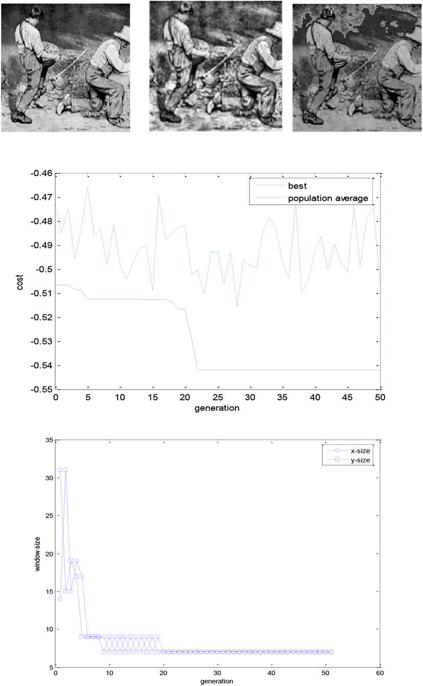
The proposed algorithm is based on Yousef’s local statistics approach [6]. The modification is the application of binary Genetic Algorithm to choose the best dimension of local masks. His algorithm is as follows:
-
Take a mask of 3×3 and slide it over each pixel one by one, from left to right and top to bottom.
-
Check any background pixel available in the mask and if available avoid that region for processing (the step is skipped in my work since there are no specific background pixels in input image).
-
At each slide of mask, calculate the mean and standard deviation of the 3×3 region of input image.
-
Compare the values of local mean and standard deviation and select the center pixel f (x, y) of mask for processing according to the following logic:
where E, k0, k1, k2 are specified parameters, ml and mg are local and global means respectively, and σl, σg are local and global standard deviations respectively
In order to apply a semi-automatic image enhancement technique, which does not fully require human intervention, and no objective parameters given by the user, a criterion for enhancement method should be chosen. The fitness function of the GA is directly related to this criterion. It is generally agreed that a good contrast and enhanced image has a high number of edgels (edge pixels). In addition to that, an enhanced image has a high intensity of the edges compared to a non-enhanced variant of the same image [11]. The number and intensity of edgels are not enough to describe a valid fitness criterion for a more naturally enhanced image. The problem is that an image can have an extreme contrast with sharp transitions from white to black and vice versa, and a relatively small number of gray levels. In this case the image will have a relatively high number of edges and a very high intensity of edges. A criterion that is proportional to number and intensities of edgels might give an oversized credit to an image that doesn’t have a natural contrast. What is needed is a quantification of the number of gray-levels present in the image, or equivalently the histogram of the image should approach the uniform distribution, as in the case of histogram equalization techniques. Taken these issues into consideration, the followed equation is suggested [1].
where F(x) denotes the fitness function applied to chromosome x, I(x) denotes the output image processed by local statistics algorithm. E(I(x)) is the intensity of the edges detected with Sobel edge detector, where the detector is applied to the transformed image I(x). n_edgels denotes the number of edgel pixels as detected with the Sobel edge detector. The term H(I(x)) is a measure of the entropy in the image I(x). H_size, V_size are the horizontal and vertical sizes (number of pixels in each direction) of the image. The Sobel detector used, is an automatic threshold detector [6].
III. EXPERIMENTATION
The GA employed has the following parameters: population size pop size=16, chromosome length nbits =16, binary tournament and K-elitism with K=4, generational type replacement, one-point crossover with selection = 0.5, mutation rate mutrate=0.15, iteration range maxit=50, k=[0.4 0.02 0.4], E=4. To demonstrate the objective performance of the proposed method, MSE and PSNR are used to compare the results. The Peak signal-to-noise ratio(PSNR)[2] is defined in decibels. Across contrast enhanced types, PSNR lacks the ability to assess image similarity. PSNR is defined as:
where maxI is the maximum value of the pixel in the image and MSE is the mean square error between the input image and the enhanced image. Hence the lesser the MSE and the bigger the PSNR, which means the better results.
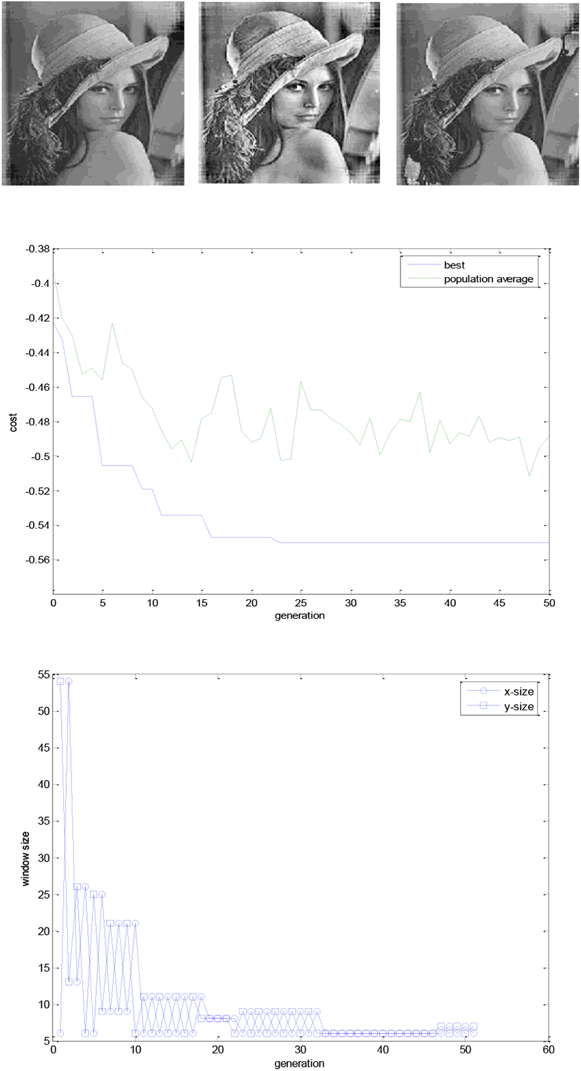
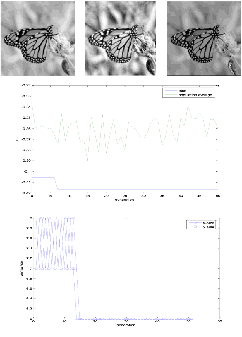
| Courbet | Lena | butterfly | ||||
|---|---|---|---|---|---|---|
| MSE | PSNR | MSE | PSNR | MSE | PSNR | |
| AHE | 57.81 | 70.33 | 31.15 | 76.52 | 96.51 | 65.21 |
| Local | 46.52 | 72.03 | 21.58 | 80.19 | 68.80 | 68.59 |
The resultant evaluation table shows that the proposed method works well compared with well-established AHE method. But the visual evaluations show that the specified parameters such as E, k0, k1, k2, may dominate the enhancement performances. Therefore, these parameters should be chosen carefully according to imaging conditions before applying GA.
V. CONCLUSION
This work show that the proposed method works successfully converge to optimal solution without human intervention. However, this preliminary experiment is not complete enough to apply in general image processing settings. The GA-generated test image sets are limited in terms of canonical examples and proper comparison approaches are missing or too simplified. Moreover, the processing time is impractically long. Further intensive research is required. In any case, the fitness of each image, taken as an individual in the population, turns out to be subjective scores given by human interpreter, but the objective fitness functions should be explored to set up for automatic GA-based processes.